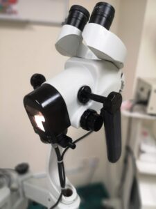
 This is a microscopic examination of the cervix, vagina, or vulva. It is used to diagnose potential abnormalities of these areas, which sometimes cannot be seen with the naked eyes. The colposcope can magnify the tissue by up to 30 times, thus making it clearer and much more accurate in terms of surface evaluation. Therefore, the biopsy of the abnormal areas performed with a colposcopic examination is more accurate than those done without the use of a colposcope.
Why do I need a colposcopy evaluation?
- It is usually recommended if you have an abnormal Pap smear (Thin Prep) test or when pre-cancerous lesion is suspected in the vagina or labia area.
Preparation for the procedure
- No specific preparation is needed. You do not need to fast.
- This procedure can be done at any time except during menstruation.

Description of the procedure
- The procedure should take about 10 to 15 minutes. There may be some discomfort but is usually tolerable. Please let your doctor know if the procedure causes significant discomfort or pain to you.
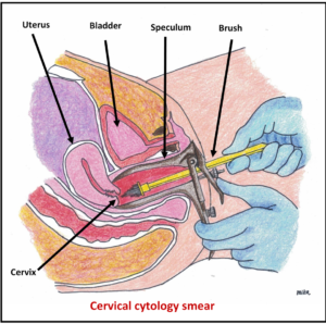
- The position for the procedure is similar to the Pap smear test.
- A speculum is inserted into the vagina to expose the cervix. The colposcope is then positioned in front of the vaginal opening to view the vagina wall and the cervix. The colposcope is usually connected to an external monitor to improve visualization. Results of the visual examination are available immediately.
- A stain or other chemical agent is applied so that the abnormal areas will become more prominent and easily seen.
- Biopsy of the abnormal area will be taken if necessary and sent for histological examination. If a biopsy is done or endocervical curettage is performed, these procedures may cause some cramping or bleeding.
Complications
There are no serious complications. A biopsy done in conjunction with a colposcopy may cause some bleeding and, rarely, an infection.
What to expect during and after Colposcopy
The colposcopy may reveal normal findings with no biopsy taken. Or it may reveal abnormal surface areas on the cervix, vagina or labia. A biopsy (small piece of tissue taken with a special forcep) will be taken from these areas. It will be sent for histological examination to look for pre-cancerous or cancerous change. The results will be available within 1 week and your doctor will discuss with you the treatment options based on this histological finding. You can read more about cervical pre-cancerous change (cervical intra-epithelial neoplasia or CIN) HERE
Post-procedure care
- If no biopsy was taken, you can resume normal activity immediately. Slight vaginal staining may occur.
- You may bathe or shower as usual.
- If a biopsy was taken, use sanitary napkins to absorb blood or drainage. Avoid sexual intercourse until the bleeding has stopped or as advised by the doctor. There is no need to restrict activities if you feel well.
- Medication is usually not necessary following this procedure.
See your doctor immediately if there is:
- Excessive vaginal bleeding which soaks more than 1 pad each hour.
- Persistent and abnormal vaginal discharge.
- Signs of infection, including headache, muscle aches, dizziness or a general ill feeling and fever.
To download a pdf copy, click HERE
[mailerlite_form form_id=3]


