Article contributed by: Dr T P Baskaran, Consultant Maternal Fetal Medicine, Obstetric & Gynaecology
Why is ultrasound used in pregnancy?
The ultrasound is the primary imaging tool in obstetric practice. It is safe at every stage of pregnancy, gives real-time images, allows detailed assessment of the structures, and helps in the monitor the fetal wellbeing. It causes no discomfort to the mother, is affordable, and the equipment is portable, making it accessible in most healthcare settings.
When are scans done during pregnancy?
In an uncomplicated pregnancy, it is recommended to have at least one good-quality scan in each trimester:
- First trimester scan: 11–14 weeks
- Second trimester scan: 18–22 weeks
- Third trimester scan: beyond 32 weeks, up to delivery
Why FAS at 18–22 weeks?
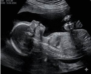
The Fetal Anomaly Scan (FAS) – also known as the Targeted Scan, Mid-Trimester Scan, Detail Scan, or TIFFA Scan – is performed during this window because the major fetal structures have completely formed and there is sufficient fluid, which allows a thorough, 360-degree view of the fetus’s physical structures or features. This timing also conveniently divides pregnancy into a pre-viable period (before 22 weeks) and a viable period (after 22 weeks), which is useful for clinical decision-making.
Patient Preparation for the FAS
- If you have a belly piercing ornament , please remove it before attending the FAS session.
- Please wear loose, comfortable clothing that allows easy access to your abdomen.
- Please arrive with a reasonably full urinary bladder, as it will may improve scan image quality.
What does the scan check?
The scan covers four main areas:
1. Growth (Biometry): Key measurements – the head circumference, the distance across the head (biparietal diameter), the abdominal circumference, and the length of the thigh bone (femur) – are taken and compared to normal growth charts for the fetal age and used to estimate the fetal weight. Additional measurements (other limb bones, parts of the brain, foot length, nasal bone length, etc.) may be taken for a more complete picture of growth. 
2. Anatomy survey: the most important and time-consuming part of the scan. The objective is to systematically survey the fetus, literally from ‘top to toe’. The extend of the ‘detail’ maybe operator dependent. However, most scan center offering this service will fulfill the minimum standards set by acceptable international guidelines. These preset guidelines, when followed will provide a comprehensive assessment of the fetus. The physical gender may be determined at this stage of the pregnancy . If you do not wish to know the gender, please inform the sonographer/ sonologist at the start of the scan.
3. Activity check: While you may simply see your baby moving on the screen, certain functional information is also recorded. Fluid in a fetus’s stomach shows they can swallow, which means their brain is functioning properly. Similarly, the filling and emptying of the fetal bladder reflects of the function of the kidney and placenta. Together with limb movements and heart activity, these observations build a picture of overall fetal well-being.
4. The baby’s environment: The scan looks at the size and position of the placenta—the vital organ that acts as your baby’s lungs and kidneys. An abnormally positioned placenta may affect how your pregnancy is managed and can carry maternal and/or fetal risks in the third trimester. The umbilical cord is examined for it attachment, the coiling pattern of its vessels, and confirmation that it has three vessels (two arteries and one vein), as well as any unusual findings such as cysts. The amount of amniotic fluid is measured (usually as the ‘Maximum Vertical Pool’, normally 2–8 cm). Any findings in the uterus or pelvis, such as fibroids, are also documented.
Scans for twins or multiple pregnancies
A scan for a single fetus usually takes 45–60 minutes. For twins or higher multiples, it can take over two hours, since each fetus needs to be assessed individually. Additional checks are also done for the placenta(s), the membranes separating the fetuses, and any differences in growth between them.

Limitation of FAS
- The fetus continues to develop throughout the pregnancy; so, some conditions which may not have been visible at 18–22 weeks and may only be detected during the third-trimester scan.
 The fetal heart is subject to some changes at soon after birth. In rare cases, if this transition does not occur normally, it will result in a heart condition. This possibility cannot be predicted before delivery.
- A genetic condition without a physical defect cannot be confirmed or ruled out by a scan alone. The classical example being the Down Syndrome fetus.
- Image quality depends on several factors: the mother’s abdominal fullness, the fetal position, the amount of fluid, and the stage of pregnancy. The quality of the ultrasound machine and the skill and experience of the operator are also crucial.
- Because of these factors, some parts of the scan may occasionally not be completed in a single visit. If a follow-up scan is advised, please do attend – for you and your baby’s best interest.
- The FAS is an important scan in every pregnancy and ideally done at the 18-22 weeks gestation
- The scope to determine the structural well-being of the fetus is wide
- It has limitations, particularly for genetic conditions, late-developing issues, and conditions that only appear after birth.
- It can be important in guiding the care plan for the rest of your pregnancy.
- Remember: A normal scan (even in the best of hands) does not always guarantee a normal baby at birth
 The fetal heart is subject to some changes at soon after birth. In rare cases, if this transition does not occur normally, it will result in a heart condition. This possibility cannot be predicted before delivery.
Key points to remember
Disclaimer
This is for informational purposes only and is not intended to be a substitute for professional medical advice, diagnosis, or treatment. It is important for readers to seek proper medical advice when necessary.